Objective: Increased computational power and improved visualization hardware have generated more opportunities for virtual reality (VR) applications in healthcare. In this study, we test the feasibility of a VR-assisted surgical navigation system for robotic-assisted radical prostatectomy.
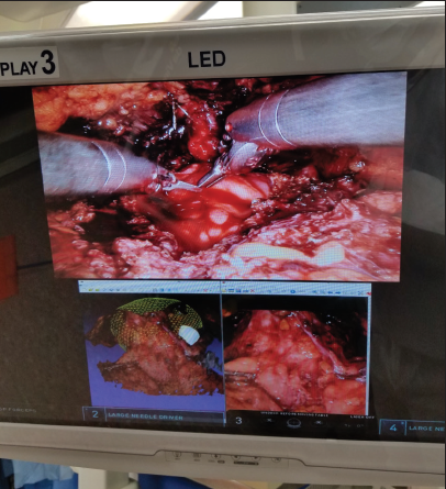
Material and methods: The prostate, all magnetic resonance imaging (MRI) visible tumors, and important anatomic structures like the neurovascular bundles, seminal vesicles, bladder, and rectum were contoured on a multiparametric MRI using an in-house segmentation software. Three-dimensional (3-D) VR models were rendered and evaluated in a side room of the operating room. While interacting with the VR platform, a real-time stereo video capture of the in situ prostate was obtained to render a second 3-D model. The MRI-based model was then overlaid on the real-time model by using an automated alignment algorithm.
Results: Ten patients were included in this study. All MRI-based VR models were examined by surgeons immediately prior to surgery and at important steps where visualization of the tumors and their proximity to surrounding anatomic structures were critical. This was mainly during the preparation of the prostatic pedicles, neurovascular plexus, the apex, and bladder neck. All participants found the system useful, especially for tumors with locally aggressive growth patterns. For small and centrally located tumors, the system was not considered beneficial due to lack of integration into the robotic console. A fully integrated system with real-time overlays within the robotic stereo viewer was found to be the ideal scenario.
Conclusion: We deployed a preliminary VR-assisted surgical navigation tool for robotic-assisted radical prostatectomies.
Cite this article as: Mehralivand S, Kolagunda A, Hammerich K, Sabarwal V, Harmon S, Sanford T, et al. A multiparametric magnetic resonance imaging-based virtual reality surgical navigation tool for robotic-assisted radical prostatectomy. Turk J Urol 2019; 45(5): 357-65.

.png)
.jpg)
.png)
.png)
