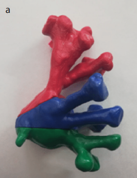
Objective: The aim of the present study was to determine the effectiveness of the use of the three-dimensional (3D) printed segmented collapsible model of the pelvicalyceal system (PCS) to improve the learning curve of the residents.
Material and methods: 3D printed models based on computed tomography (CT) images of 10 patients with a staghorn stone were developed. Used images of patients were obtained from CT scans in Digital Imaging and Communications in Medicine format. The area of interest was extracted and saved in stereolithography format. Further, the bioengineer performed virtual segmentation corresponding to the level of each calyces group that was defined by an experienced urologist. The final stage was the printing of each separated detail. Special questionnaire for evaluating the effectiveness of 3D models during both the examination of PCS anatomy and planning percutaneous nephrolithotomy was invented.
Results: The determination of the anterior and posterior calyces of the upper group was improved by 61% and 69%, the difference in the determination of the calyces of the middle group was 60% and 51%, and the answers regarding the number of the anterior and posterior calyces of the lower group became better by 67% and 74%, respectively (p<0.001). The ability to select the optimal calyx for the primary and the second access became better by 60% and 55%, respectively (p<0.001).
Conclusion: 3D printed segmented collapsible model of the PCS is promising for the improvement of the learning curve of residents and enables to optimize their clinical education, as well as compensate for their lack of experience.
Cite this article as: Guliev B, Komyakov B, Talyshinskii A. The use of the three-dimensional printed segmented collapsible model of the pelvicalyceal system to improve residents’ learning curve. Turk J Urol 2020; 46(3): 226-30.

.png)
.jpg)
.png)
.png)
